


Salpingography Set
A Salpingography set includes several key components such as a catheter, contrast medium, and radiologic equipment, all of which are crucial for the successful diagnosis of fallopian tube patency and uterine abnormalities. The procedure provides important insights into a woman’s reproductive health, especially for those struggling with infertility. While the procedure is generally safe, it is important for healthcare professionals to follow all sterilization and safety protocols to minimize risks and ensure accurate results.
Description
Salpingography is a radiographic procedure used to assess the fallopian tubes and the uterus in women, primarily to diagnose infertility, tubal blockages, or other uterine and tubal abnormalities. The procedure involves injecting a contrast dye into the uterus and fallopian tubes, followed by an X-ray to visualize the passage of the dye. The set used for this procedure is specifically designed to ensure both accuracy and safety.
Components of a Salpingography Set
A typical Salpingography set contains several components required for the procedure, each serving a specific purpose in ensuring proper technique and patient safety.
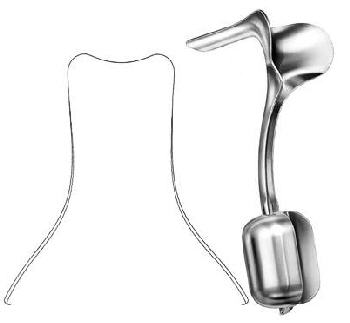
- Catheter (Intrauterine Catheter)
- Description: A flexible, sterile tube inserted into the cervix to deliver the contrast dye into the uterus. It must be small enough to be safely inserted yet durable to handle the pressure of the dye being introduced.
- Material: Usually made of medical-grade silicone or plastic, ensuring flexibility and minimal discomfort.
- Size and Design: The catheter has a tapered design for smooth insertion. The end of the catheter may be designed to create a seal in the cervical canal to prevent leakage of the contrast medium.
- Contrast Medium (Dye)
- Description: A radiopaque substance used to make the fallopian tubes visible on an X-ray. This dye should be water-soluble, non-toxic, and should have good visibility on X-ray.
- Common Contrast Agents: Examples include a water-based solution like iodized contrast media (e.g., Hysterosalpingography solution), which is often used in the procedure.
- Function: The contrast medium will flow through the uterus and fallopian tubes. Any blockage or irregularities in the tubes will be visible as the dye outlines the contours of the reproductive organs.
- Syringe and Injector
- Description: A sterile syringe used to inject the contrast medium through the catheter into the uterus and fallopian tubes.
- Size and Material: The syringe typically ranges from 10-20 mL and is made of medical-grade plastic. It is important to ensure that the syringe is capable of withstanding pressure when injecting the contrast medium.
- Usage: The physician or radiologist injects the dye slowly to allow the X-ray technician to capture the images at the right moments.
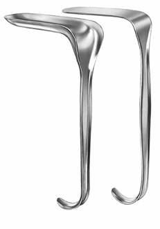
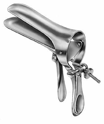
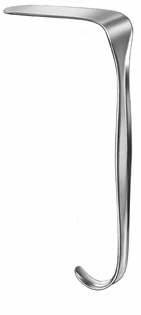
- Speculum
- Description: A device used to open the vaginal walls to gain access to the cervix. It is used to visualize and insert the catheter into the cervix.
- Material: Typically made of metal or plastic.
- Size: Available in different sizes depending on the patient’s anatomy.
- Radiology Table and Imaging Equipment
- Description: The patient is usually positioned on an X-ray table. The radiographic machine (fluoroscopy or conventional X-ray) is used to capture the images of the uterus and fallopian tubes after the contrast medium is injected.
- X-ray Machine: Often includes a fluoroscopic unit with real-time imaging capabilities. It helps to visualize the flow of the contrast medium immediately as it fills the uterus and fallopian tubes.
- Sterile Drape and Gloves
- Description: Sterile equipment to maintain a hygienic environment during the procedure. Gloves are worn by the healthcare provider to ensure infection control, and a sterile drape is used to cover the area around the cervix.
- Purpose: These ensure that the procedure is carried out in a sterile environment, reducing the risk of infection.
- Antiseptic Solution
- Description: An antiseptic solution, such as iodine-based or chlorhexidine-based solutions, is used to cleanse the external genitalia and cervix before the procedure to prevent infection.
- Application: The area around the cervix is cleaned thoroughly to avoid contamination during catheter insertion.
- Additional Diagnostic Tools (Optional)
- Description: In some cases, additional diagnostic tools may be used to assess the uterus and fallopian tubes further, especially if abnormalities are detected during the procedure.
- Examples: This may include a uterine sound (to measure the depth of the uterus), biopsy instruments (if necessary), or saline solution (for a saline infusion sonography).
Procedure Overview
- Preparation:
- The patient is asked to empty her bladder, and a sterile field is created.
- A speculum is inserted into the vagina to expose the cervix.
- The cervix and vaginal area are cleaned with an antiseptic solution.
- Catheter Insertion:
- A catheter is gently inserted through the cervix into the uterus.
- The catheter is positioned in such a way that the contrast dye will flow through the uterine cavity and into the fallopian tubes.
- Injection of Contrast Medium:
- Once the catheter is in place, the contrast medium is injected into the uterus and fallopian tubes.
- The radiologist or technician monitors the flow of the dye, looking for any obstructions or abnormalities in the fallopian tubes.
- X-ray images are taken during the process to visualize the spread of the dye.
- Imaging:
- X-ray images are captured at different angles to get clear and comprehensive views of the uterus and fallopian tubes.
- A blockage in one or both fallopian tubes will prevent the contrast medium from passing, indicating a tubal obstruction.
- Post-procedure Care:
- Once the procedure is complete, the catheter is removed.
- The patient may experience some mild cramping or spotting, but this usually resolves quickly.
- The results of the salpingography will be reviewed, and further diagnostic procedures or treatments may be recommended based on the findings.
Applications of Salpingography
- Infertility Evaluation: One of the primary uses of salpingography is to evaluate the fallopian tubes in women experiencing infertility. It can help diagnose conditions like tubal blockages, adhesions, or other abnormalities.
- Diagnosis of Ectopic Pregnancy: The procedure can also detect if there has been a prior ectopic pregnancy that may have caused scarring in the tubes.
- Evaluation of Uterine Abnormalities: Salpingography can help identify uterine problems such as fibroids, polyps, or congenital anomalies like septate uterus, which may contribute to fertility issues.
- Tubal Patency Test: It is an effective method to test if the fallopian tubes are open and functioning, which is essential for conception.
Advantages and Limitations
Advantages:
- High Diagnostic Value: Provides clear and detailed images of the fallopian tubes, making it an essential diagnostic tool.
- Minimally Invasive: It is less invasive than surgical alternatives for diagnosing tubal patency.
- Procedure Speed: The procedure is relatively quick, usually taking between 20-30 minutes.
Limitations:
- Radiation Exposure: Though the radiation dose is minimal, it is still a concern, especially in women who may undergo multiple procedures.
- Discomfort or Pain: Some women may experience mild to moderate discomfort during the procedure, including cramping and spotting.
- Risk of Infection: Though rare, there is a small risk of infection due to the insertion of the catheter.
Reviews
There are no reviews yet.